ADMdx Logo

CorInsights® MRI – See clearly what IMAGING INSIGHTS can reveal for patient care 

CorInsights® MRI is our FDA-cleared diagnostic report that provides exceptional insights into brain volumes. Numerous published studies have shown that various brain conditions, such as Alzheimer’s disease, are often associated with volume loss in certain brain region combinations.  Using a standard MRI scan, our CorInsights® MRI report quantifies which, if any, brain regions have reduced volume compared to an age- and sex-matched normative reference. It translates complexity to clarity by integrating this information into a clear overall view of effects.  This insight is intended to aid the medical professional as an adjunct to clinical assessment.

Quantitative accuracy transformed to integrated insight

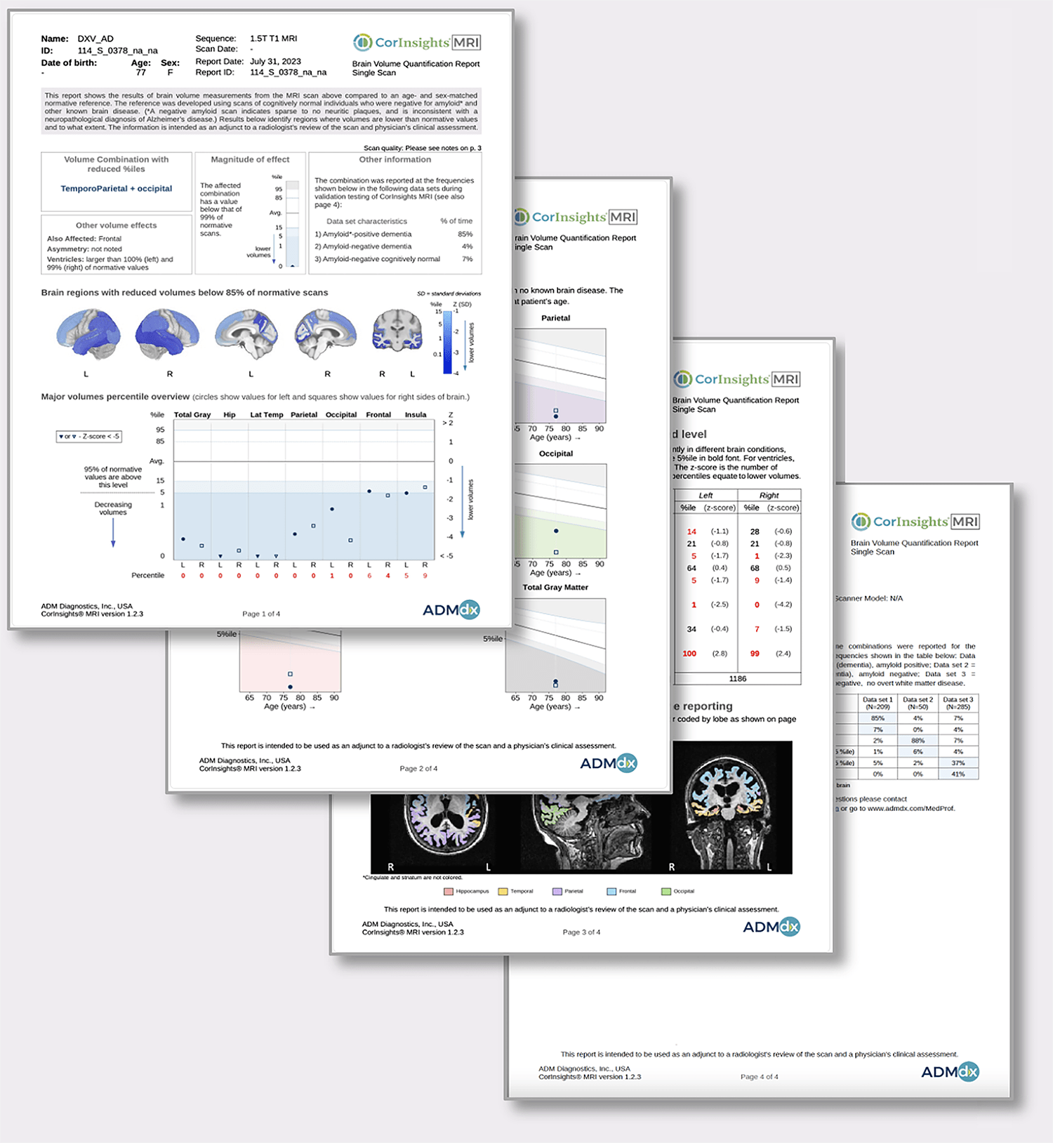
CorInsights® MRI provides an illustrated, quantitative report to aid in the assessment of patient brain status.  CorInsights® MRI:

  • Measures brain volumes and determines percentiles (amount of difference) compared to age- and sex-matched normative values
  • Identifies the combination of regions that represents the lowest percentiles below the reference range
  • Translates these results into cohesive images and graphs that can be readily interpreted by the medical professional
  • Identifies the volume combination, or pattern, that is most affected
  • Provides patient-friendly graphs that can be shared
  • Requires only a T1-weighted MRI scan for which reimbursement is available

Integrated insights offer greater clarity

Feedback from neurologists, gerontologists, and other medical professionals who have evaluated CorInsights® MRI

  • “There’s a real need for this.”
  • “I think the reports are excellent.”
  • “This will definitely help with earlier and more accurate diagnosis…”
  • “This is amazing!  I’m going to show my colleagues!”
  • “For me the first step in my clinic has always been to look for clear evidence of neurodegeneration. Once we rule that out, we can focus on a whole bunch of different approaches. That's why the CorInsights® is so important for me.”

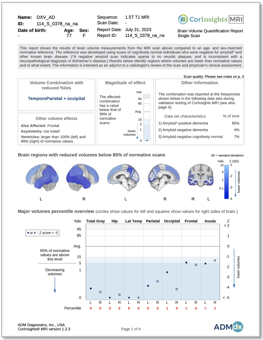
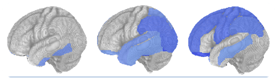
CorInsights® MRI shows the overall picture as well as the detailed quantitative measures that support findings.  Shown at left, see on page 1 the overall pattern of effect as a color-coded image and by name, as well as percentile plot and information regarding other attributes.

Easy to order. Easy to understand.

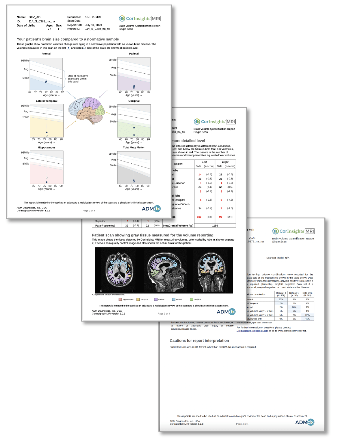
On page 2 of the reports, find patient-friendly aging curves, showing how their volumes compare to the normative reference in major regions

On page 3, review the segmentation of major lobes, and see percentile and z-core results for the subvolumes that are color-coded in the image on page 1

On page 4, see additional quality control information and any cautions noted during CorInsights® MRI measurement, as well as a summary of methods

Support your entire team:

CorInsights® MRI makes patient care more effective, while providing important information to patients and their caregivers

Radiology
Receives quantitative visualization of the individual structures and patterns of structures affected by low volumes, with efficient radiology workflow integration

Patient-facing medical professional
Gains clarity and increased confidence based on the neuroimaging specialist’s intepretations, and gains tools that can be used to convey findings to the patient

Healthcare network
Using a reimbursed scan and quantitation CPT code, your entire healthcare network gains access to enhanced tools to optimally serve patients and staff

Patient and caregiver
Gains increased clarity, understandable results, and increased confidence through their medical professional’s assessment

Benefit from the insights of brain 
volume quantitation

Numerous published studies have shown that various brain conditions, including different types of dementia, are associated with accelerated brain volume loss (atrophy) in certain brain regions, which can be quantitated from an MRI scan. For example: Alzheimer’s disease is often associated with progressive Temporal and Parietal volume decreases1-3. FrontoTemporal dementias are often associated with loss in certain frontal and temporal lobe regions4-5.

Brain volume quantitation can provide a valuable tool when combined with other assessments

Gain confidence from our
extensive validation

Volumes measured by CorInsights® MRI have been tested using cognitively normal, amyloid-negative subject scans, as well as data sets expected to have volumes in certain region combinations below the normative range based upon well-established literature. Our data sets include individuals with amyloid status confirmation and clinical diagnoses of:

  • Mild Cognitive Impairment

  • Late Onset Alzheimer's disease

  • Early Onset Alzheimer's disease

  • FrontoTemporal Dementia: Behavioral

  • FrontoTemporal Dementia: Semantic

  • Posterior Cortical Atrophy

  • Other conditions

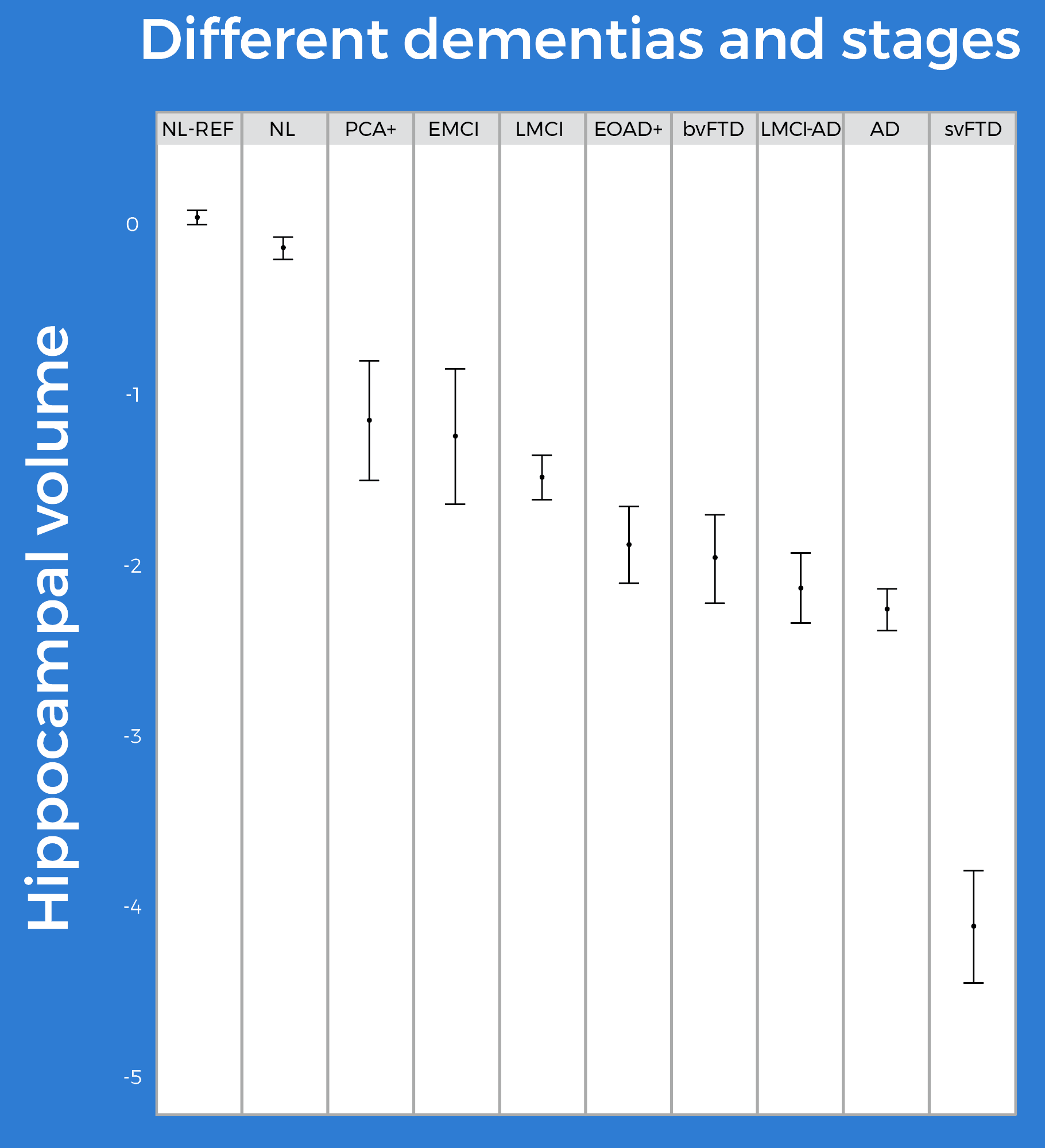
CorInsights® MRI values were compared to the expected percentile and z-score ranges for these data sets and were confirmed to be in these ranges. Relationships between regions were also found to be in agreement with published literature.

Our quantitation starts with quality

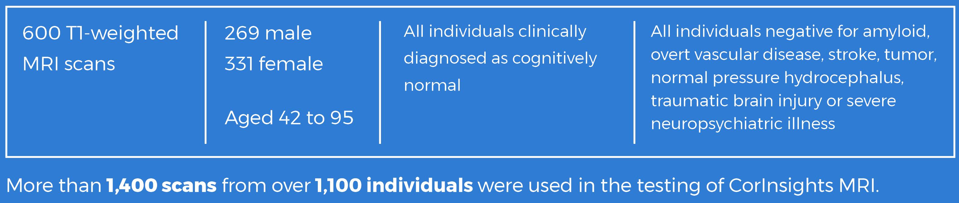
In the development of the CorInsights® MRI normative reference database, data were extensively screened to ensure that CorInsights® MRI normative values are not influenced by disease, and consisted of:

REMARKABLE CLARITY is the result

ADMdx Logo
PRIVACYCONTACT
©2023. CorInsights® MRI is a registered trademark of AMDdx, 555 Skokie Blvd, Suite 500, Northbrook, IL, 60062, USA.